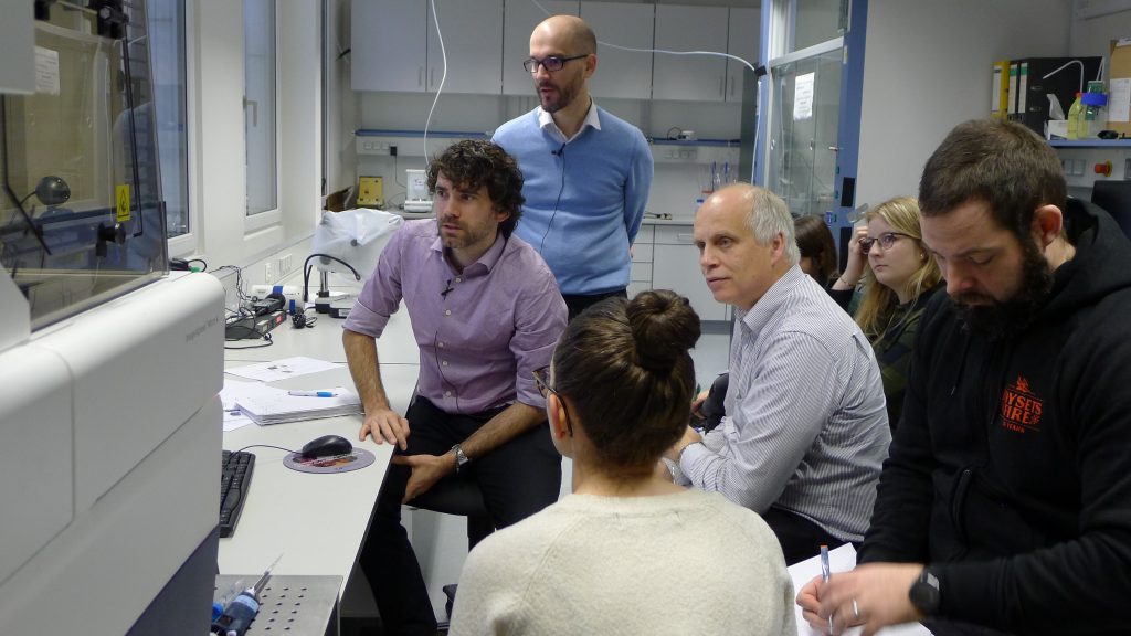
A state-of-the-art fluorescence microscope, which Kaiserslautern University of Applied Sciences has just acquired, promises deep insights into the respective objects of study. The 500,000 euro device is located at the Zweibrücken campus. It was funded by the German Research Foundation, among others.
“With our fluorescence microscope, we now have the opportunity at the Zweibrücken campus to carry out high-throughput screening as we know it from the pharmaceutical industry,” says Prof. Bernd Bufe from the Chair of Molecular Immunology and Immunosensors. The newly acquired large device consists of a microscope and an environmental chamber in which living cells can be examined. This enables cells, organisms or tissue to be stimulated with various chemicals. A pipetting robot makes the examination process much faster. Prof. Bufe will use the device to conduct further research into the immune-odor interaction. Prof. Karl-Herbert Schäfer, responsible for biotechnology in microsystems technology, will be investigating questions relating to the enteric nervous system of the intestine, while Prof. Tanja Brigadski from the Chair of Optical and Electrophysiological Analysis Methods in Biomedicine will be working on the topic of growth factors and learning processes in neurons. Collaborations are also planned outside the university. For example, Prof. Markus Bischoff from the Institute of Medical Microbiology and Hygiene at Homburg University Hospital wants to research healing processes after tissue injuries. “We have a device here that is particularly suitable for short-term experiments lasting from milliseconds to an hour,” says Prof. Bernd Bufe, describing the new acquisition. The microscope enables the researchers to recognize even the tiniest structures. “We have a penetration depth of up to six cell layers,” explains Prof. Bernd Bufe, “which allows us to see small organoids that work like mini-brains in the digestive tract, for example. We can literally watch the gut think.” Even 3D images of living cells are no problem for the microscope. “We are able to look deep into a cell and make optical sections right through a living cell. This gives us new opportunities to examine entire cell clusters,” says the professor happily. The device is fully automated and produces images ready for publication. Naturally, this generates a lot of data. This is why the university is also building a high-performance IT infrastructure.
Anyone interested in using the device for complex questions on biological processes is welcome. Contact: Prof. Bernd Bufe, phone: 0631 3724 – 5410, e-mail bernd.bufe(at)hs-kl.de
